There are diseases that can, within a very short time, bring a healthy person to a state with extremely unfavorable prognosis. One of them is Lyell's syndrome (toxic epidermal necrolysis). It begins with a sharp deterioration in health, the patient’s skin peels off, hair falls out, and internal organs fail one after another. The pathology was first described in 1956 by Scottish dermatologist A. Lyell. He personally observed four similar cases, then, on his initiative, a survey of all doctors practicing in the UK was carried out. It was possible to collect 128 episodes and use them to study in detail the mechanisms of disease development.
Etiology and pathogenesis
Since 1970, annual statistics on the spread of Lyell's syndrome have been compiled, which has made it possible to highlight the frequency of the disease. It occurs in 1.3% of cases per million population per year. Mortality is high: 60% of patients with this diagnosis die.
Discussions about the causes of the origin of the syndrome and the mechanisms of its development are still ongoing. So far, only one form of pathology has been well studied - medicinal. It develops after the formation of disturbances in the human body’s ability to neutralize intermediate reactive metabolites that accumulate in cells after taking certain medications. They, reacting with tissue metabolites, form an antigenic complex that causes a certain immune response. So it gives impetus to the development of an allergic reaction.
Toxins produced in large quantities during the decay of microbes can also provoke the formation of an antigenic complex. Due to malfunctions in the neutralization system, they are not eliminated naturally; as a result, antigens combine with proteins of the epidermal cell. The immune system perceives it as a “stranger” and turns on a protective mode aimed at destroying it. As a result, a reaction develops that leads to necrosis of epidermal cells.
The pathogenesis can be compared to donor organ rejection syndrome, which develops due to incompatibility. The role of foreign tissue (in our case) is played by the patient’s own skin. The most likely provoking factor is considered to be an acute infectious process associated with infection with staphylococci.
At-risk groups
Today, a genetic predisposition to Lyell's syndrome has been proven. Scientists believe that the presence of a chronic source of infection (tonsillitis, sinusitis, cholecystitis) in the human body increases the risk of disease. Women are more often affected; no direct relationship between the increase in the incidence of pathology and the age of patients has been identified.
Anyone who suffered Lyell's syndrome at an early age is at risk. In children, in most cases, the disease has an infectious etiology. The trigger point for its development is associated with the course of acute respiratory viral infection and the therapy received for this reason using non-steroidal anti-inflammatory drugs. The second most common provoking factor is damage to the body by staphylococci.

In adults, those at risk are those who constantly experience nervous overload, psychological stress, who have been depressed for a long time, who self-medicate and drink various medications without control. You should not take sulfonamides, antibiotics, anticonvulsants, anti-inflammatory and painkillers, vitamins and contrast agents used in radiography without a prescription. You should be careful when choosing dietary supplements, immunosuppressants and immunomodulators.
A special risk group includes people with HIV status. Their incidence rate is a thousand times higher than in the general population.
Causes of pathology

Modern medicine identifies several variants of the disease, which depend on the root cause. The first is an allergy to an infectious process, often caused by the activity of Staphylococcus aureus. The pathology most often occurs in children and is characterized by a severe course.
The second option is malignant pemphigus, which appeared due to the use of certain medications (antibiotics, sulfonamides, acetylsalicylic acid, anticonvulsants, antituberculosis, anti-inflammatory and painkillers). Often the development of such a syndrome is associated with the simultaneous use of several medications. Recently, there have been cases of the appearance of the syndrome due to the use of vitamin complexes, dietary supplements, radiography, etc.
Forms and types of disease
Experts, taking into account the different etiologies of the pathology, have identified the following forms of the disease:
- The drug develops as a result of drug exposure.
- Staphylogenic occurs as a complication of a bacterial infection. This form develops only in children. It does not depend on the use of drugs and is not fatal. The prognosis for such a diagnosis is always favorable.
- Idiopathic form. It includes all cases in which it is not possible to identify the trigger points of the disease.
- A form that occurs with secondary pathologies. This group includes those episodes in which Lyell's syndrome develops against the background of psoriasis, chickenpox, herpes zoster or pemphigus.
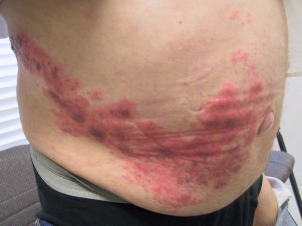
The acute course of any form has the same clinical picture.
Symptoms and signs
The disease starts suddenly, the patient’s condition constantly worsens, and death can occur in a short time (5-6 hours). That is why it is so important to know what it is - toxic necrolysis of epidermal tissue.
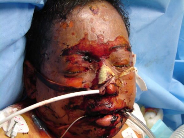
Initially, a sharp hyperthermia of the skin is recorded. Its temperature rises to the highest possible temperatures, and single red spots form on the surface of the inflamed area, which quickly turn brown. They are surrounded by edematous tissue. The affected elements quickly merge into one large spot; any touch to it causes severe pain.
Then, in place of the red spots, a huge number of large (palm-sized) blisters appear, filled with serous fluid. Outwardly, they strongly resemble burn blisters. After spontaneous opening, bleeding ulcers, similar to erosions, remain in their place. If the inflamed skin is touched, its epidermal layer will begin to peel off.

In parallel with the damage to the skin, changes in the mucous membranes are observed. They become covered with cracks and abrasions, causing severe pain. Even with a light touch, areas with broken integrity begin to bleed. Eating becomes difficult.
The described reaction can also affect the inner lining of the nasopharynx, bronchi, gastrointestinal tract, and urethra. As the disease progresses, such conditions cause irreversible consequences, so the mortality rate is very high.
Damage to the skin and mucous membranes is accompanied by unbearable headaches and excruciating thirst. Over time, dehydration occurs, the patient becomes lethargic, and the blood thickens. This also complicates the operation of all internal systems. As a result of necrosis of the epidermis, toxins are formed and have a negative effect on the brain. Open wounds on the body give impetus to the development of purulent inflammatory processes, blockage of blood vessels occurs, which causes circulatory problems. That is why it is so important to provide medical care to the patient when the first symptoms of illness appear.
Mechanism of disease development
Great importance in the development of pathology is assigned to heredity to allergies. In the clinical picture of a large number of patients there are indications of diseases of an allergic nature:
- allergic asthma;
- dermatitis;
- hay fever;
- rhinitis, etc.
A protein in the skin of such patients combines with the introduced drug substance, forming an antigen for Lyell's symptom. This is how human immunity not only reacts to the administered medication, but also to the patient’s skin. This process is similar to the body’s rejection of an implant, in which the immune system perceives the patient’s skin as the implant.
The basis of the syndrome is the so-called Sanarelli-Schwartzman phenomenon, which is a reaction of the immune system leading to failures in the regulation of protein breakdown and the accumulation of breakdown products in the body. As a result, internal systems and organs suffer from damage from toxins. This, in turn, disrupts the functioning of the organs responsible for toxic neutralization, which aggravates the situation, leading to changes in electrolyte and water-salt balance. These processes worsen the patient's condition in the presence of Lyell's syndrome and can easily lead to his death.
Characteristic symptoms of the syndrome
The onset of the disease is accompanied by an uncontrolled and spontaneous increase in temperature up to 40 degrees. Within two to three hours, painful formations appear on the skin, which may merge slightly.
After twelve to fifteen hours, peeling of the epidermal layer begins in areas of seemingly healthy skin. This process is accompanied by the formation of bubbles of various sizes. After their opening, noticeable erosions remain on the skin, surrounded by edematous skin. A bloody substance is released from the blisters, which leads to severe dehydration of the body .
In the presence of the syndrome in question, peeling of the skin is also observed in response to a minor impact on its surface. The skin quickly turns red and hurts on palpation. Sometimes all this can be accompanied by a profuse rash. In young children, Lyell's disease often begins with signs of conjunctivitis .
Diagnostics
When confirming the diagnosis, a correct assessment of the existing clinic and the area of skin lesions is of great importance. Depending on this indicator, differential diagnosis of Lyell's syndrome from Steven-Johnson syndrome is carried out.
Laboratory blood tests are required. With the described pathology, the following is found:
- low eosinophil count or their absolute absence;
- decrease in the number of leukocytes;
- increase in band and neutrophils;
- low protein levels;
- increase in globulins.
Liver tests show elevated values of all existing standards; kidney tests can detect high levels of urea. Electrolyte balance is disturbed. To clarify the diagnosis, a biopsy of the exfoliated area of skin may be performed. It should confirm the peeling of the entire epidermis. For comparison: with “burnt skin” syndrome, only the stratum corneum peels off.

If a drug is suspected, the doctor may order an immunological test. With its help, it is possible to find a medication, the use of which provoked the development of such an allergic reaction. It is important to exclude it from the medical protocol when drawing up a therapeutic regimen.
Basic diagnostic techniques
A general blood test in the presence of a disease allows you to identify the inflammatory and infectious process. The absence or decrease in the number of eosinophils in the blood is a sign that makes it possible to distinguish other allergic reactions from Lyell's syndrome. Data obtained during a coagulogram may indicate excessive blood clotting. Blood biochemistry and urinary analysis make it possible to detect malfunctions in the kidneys and monitor the patient’s condition during therapy.
One of the main tasks of diagnosis is to determine the drug that provoked the reaction, because its repeated use can lead to death of the patient. The provoking substance can be identified using immunological testing. The causative drug may be indicated by intensive proliferation of immune cells, which appears due to its introduction into a blood sample that was previously taken from the patient.
Histological examination and biopsy of the skin make it possible to learn about the condition of the superficial epidermal cells. In the depths of the tissues, the formation of vesicles, swelling and mass localization of immune cells can be observed.
Treatment methods
It is impossible to fight Lyell's syndrome on your own; all necessary measures can only be carried out in a hospital setting, in intensive care wards or in burn centers. The patient must first be completely undressed and placed on a special bed, which is capable of blowing the patient’s body using jets that release sterile air. The first goal of therapy is to relieve pain, eliminate increased sensitivity of the skin, and stop the development of a toxicological reaction. For this purpose the following is carried out:
- Cascade plasma filtration (blood purification). If it is not technically possible to perform it, hemosorption is used. The sooner these procedures are performed, the better the prognosis. If they are carried out within the first two days from the onset of the disease, a rapid recovery is possible.
- Massive systemic therapy with drugs from the group of corticosteroids. Dosage regimens are developed on a case-by-case basis, taking into account the patient’s age and weight.
- Administration of intravenous immunoglobulins. The use of such agents can reduce the incidence of bacterial complications and prevent the development of the pathological process.
- Hyperbaric oxygenation. This procedure helps increase the oxygen capacity of the blood.
Every 72 hours, the patient must be examined by doctors whose specialization involves the treatment of diseases associated with damage to the mucous membranes.
To prevent dehydration, the patient must be fed constantly. He can be given warm water in which powder products that can restore rehydration (Regidron, Oralit) have been previously dissolved.
Local therapy
Treatment of the skin is carried out according to the principle of burn therapy. Sterile dressings are applied to the erosive surfaces; they are pre-treated with creams and ointments that contain hormones and antibiotics (Methylprednisolone, Contrical, Prednisolone).
It is important to understand that the disease cannot be treated with folk remedies. Any such initiative can end in complications. The most common of them is the addition of a bacterial component. It significantly worsens the patient's condition.

The incidence of pneumonia, pyoderma and sepsis is high. Almost always, the course of the disease is accompanied by deterioration in cardiac activity and the appearance of respiratory failure. In severe cases, kidney function may stop working and, as a result, death from toxin poisoning may occur. Dehydration leads to increased blood thickness. This phenomenon promotes the formation of blood clots in the pulmonary and cardiac systems. And this is also instant death.
Violation of the integrity of the mucous membrane of internal organs is associated with the risk of extensive internal bleeding. When the conjunctiva of the eyes is damaged, the development of blindness becomes possible. After therapy, it disappears, but the patient remains photophobic for a long time.
Lyell's syndrome - description
Lyell's syndrome (toxic epidermal necrolysis) is the most severe variant of allergic bullous dermatitis. Named after Scottish dermatologist Alan Lyell (1917–2007), who first made a detailed description of this disease in 1956.
Most often, Lyell's syndrome is a reaction to drugs (antibiotics, sulfonamides, NSAIDs).
Story
This pathology was first described in 1956 by Scottish dermatologist Alan Lyell. He systematized 11 reports of 14 cases of this disease, 4 of which he personally observed.
Lyell replaced the previously accepted designation “acute pemphigus” with “toxic epidermal necrolysis” (TEN). The fact is that all patients o. The rash served as the main diagnostic sign for identifying a new nosological unit. Lyell coined the term “toxic” because he believed that the disease was caused by toxemia, a specific toxin circulating in the body. “Necrolysis” is a medical neologism invented by Lyell himself, who combined in it the main clinical sign - “epidermolysis” and the histopathological sign - “necrosis”.
Lyell also noted the severity of damage to the mucous membranes and the unusual weakness of inflammatory processes in the dermis - “dermal silence.”
In the scientific world, the disease received the name of its discoverer during Lyell’s lifetime - in the 60s. However, the dermatologist himself always used the designation TEN.
In 1967, after conducting a targeted survey of colleagues throughout the UK, Lyell presented the most extensive summary of TEN to date - 128 cases of the disease. In the same year, he described 4 forms of TEN, corresponding to a specific etiology: staphylococcal, drug, mixed and idiopathic.
Currently, the staphylococcal form of Lyell's syndrome is identified as a separate nosological unit: staphylococcal scalded skin syndrome (SSSS, L00 according to ICD-10).
Prevention methods
There is no specific prevention yet, the disease itself has not been fully studied, and all the factors that can trigger the onset of a complex systemic allergic reaction are unknown. Experts advise everyone, without exception, to take medications only as prescribed, strictly follow the prescribed dosages, and each time inform your doctor about adverse reactions that occur as a result of the medications used. You cannot take more than five different drugs at the same time. Self-medication of people who are predisposed to allergies is considered unacceptable.
Prevention measures
Every parent should know preventive measures to prevent the occurrence of allergic reactions in the child’s body, and especially Lyell’s syndrome. You need to follow simple rules, they will help keep your baby healthy:
- do not self-medicate your child, take only medications prescribed by a doctor;
- strengthen the baby’s body’s defenses from birth;
- if strange reactions appear on the skin, do not wait for the disease to develop, but seek help from specialists;
- every parent should know and remember what medications the child is allergic to, especially antibiotics;
- When prescribing medications by a pediatrician, always pronounce the name of the allergic substance without waiting for a question from the doctor;
- do not use medications that have expired or were improperly stored in treating a child;
- An antihistamine should always be in your home medicine cabinet.
Simple preventive measures will help to avoid Lyell's syndrome and maintain the health of not only children, but the entire family. Lyell's syndrome should not be considered a death sentence. If a person takes first aid measures in time and turns to specialists as soon as possible, the chances of a complete recovery of the body are high.
Forecast
If the diagnosis is confirmed, the prognosis is always serious. As a percentage, the mortality rate ranges from 35-70%. Much depends on what prognostic factors can be identified in each individual patient. In the West today, a scale has been developed by which the severity of the condition can be calculated and accurate predictions made. The patient’s age, heart rate, the presence or absence of cancer, the area of the affected surface, the level of urea in the blood, the level of plasma bicarbonates and glucose are taken into account.
The highest mortality rates are in younger age groups, in older people, and in patients with a history of immunodeficiency states. The patient dies from complications.
